
Fig. 6.
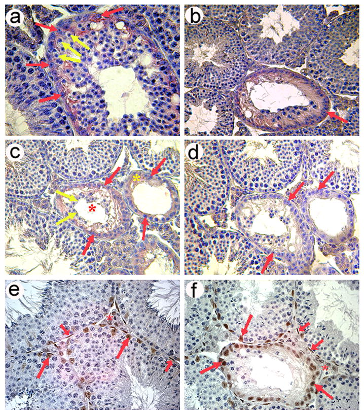
Degenerating seminiferous tubules in Foxa3-deficient testes re-express Anti-Muellerian hormone (AMH) as revealed by immunohistochemistry (a–d). a) A seminiferous tubule in the early phase of degeneration. The thickness of the germinal epithelium appears to be normal, although vacuolization of the epithelium has already occurred. Germ cells present in this tubule are mostly round spermatids as judged from nuclear morphology. Earlier germ cell stages (spermatogonia and spermatocytes) are rather rare. Germ cells are devoid of any pink stain (yellow arrows) but the surrounding cytoplasm of the Sertoli cells is clearly stained (red arrows). Tubules with normal appearance do not exhibit any staining (lower left part). b) A degenerating tubule in a more advanced stage than that shown in a). One generation of spermatocytes (presumably maturing to spermiation) is still present in this tubule. Sertoli cell nuclei have aligned along the tubular wall and the cytoplasm is clearly stained by the AMH antibody (red arrow) c) A tubule during degeneration (red asterisk) still containing some germ cells (yellow arrows) and a tubule at the final stage of degeneration (yellow asterisk), which exhibits a Sertoli cell only-phenotype characterized by the complete loss of germ cells. At this stage the Sertoli cells have flattened and extend little beyond their nuclei. Again, AMH immunoreactivity is widespread. Red arrows point out AMH positive Sertoli cells. d) Negative control omitting the AMH antibody from the diluent. Also, the adult wild type control mice exhibited no staining while Sertoli cells in young testes up to p10 showed strong cytoplasmic Sertoli cell staining (data not shown). e) Expression of the androgen receptor (AR; brown stain) in a wt testis as revealed by immunohistochemistry. Sertoli cells (long red arrows), peritubular myoid cells (short red arrows) and Leydig cells (red asterisk) are AR positive. f) AR expression in an 8 months old mutant testis showing staining in the same cell types as the age-matched wildtype control. Remarkably, also Sertoli cells in almost completely degenerated tubules still express the AR.