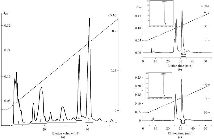
Figure 1.
Isolation of the proteins. (a) Separation of crude V. nikolskii venom by cation-exchange chromatography on HEMA BIO 1000 CM column (8 × 250 mm) in an ammonium acetate gradient from 5 mM to 1 M in 100 min. Flow rate 1 ml min−1. (b) and (c) Isolation of proteins VN4-3 and VN5-3 by reverse-phase HPLC on a Vydac C18 column (10 × 250 mm). The MALDI mass spectra for VN4-3 and VN5-3 are shown in insets. The dashed lines show the gradients of ammonium acetate (in a) or acetonitrile (in b and c); C are the concentrations of ammonium acetate (a) or acetonitrile (b and c).

