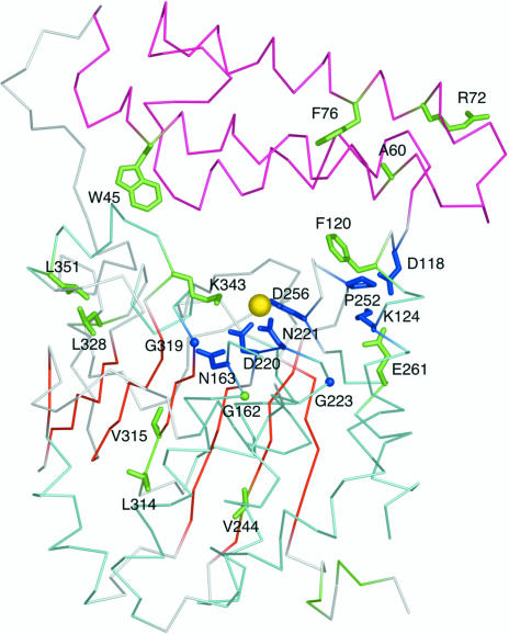
Figure 3.
Fully (blue) and highly conserved (green) residues of the Pfam01937.11 family mapped onto a Cα trace of the At2g17340 structure. The color coding of the structure is consistent with that introduced in Fig. 1 ▶. The yellow sphere represents a putative Mg2+ ion coordinated within the protein. The clustering of conserved amino acids around the metal atom and in the cleft between the amino-terminal three-helix cluster (pink) and the carboxy-terminal domain (cyan and red) suggests the location of the active site. The figure was generated using PyMol (DeLano, 2002 ▶).

