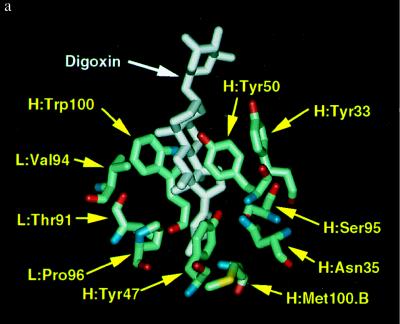
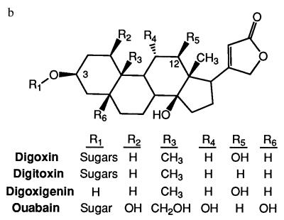
Figure 1.
(a) Computer model showing bound digoxin and 10 residues that define the 26–10 binding pocket. This figure was generated using Quanta charmm software (Micron Separations) using the coordinates of Jeffery et al. (22). (b) Structures of digoxin and the three analogs used in these studies.


