
Figure 2.
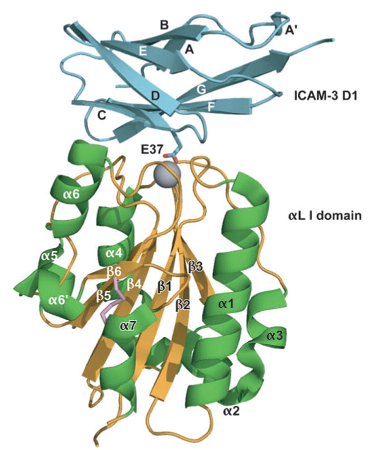
A mutant, high-affinity αL I domain (gold β-sheet and coil and green α-helices) in complex with domain 1 of ICAM-3 (cyan). The Mg2+ is shown as a gray sphere. The side chain of the key integrin-binding residue, Glu37 of ICAM-3, is shown. The mutationally introduced K287C/K294C disulfide bond that stabilizes the open conformation is shown in pink. ICAM-3 domain 2 is omitted for clarity. [From Protein Data Bank (PDB) ID code 1T0P (7).]