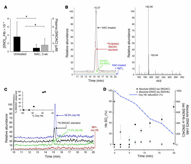
Figure 3. SNOAC is formed from NAC in blood ex vivo and in vivo.
(A) The SNOrbc in heparinized LV blood (black bars), measured by reductive chemiluminescence (11), was lower than normal following 3 weeks of treatment with 10 mg/ml NAC (n = 3–4 each). In the same mice, plasma SNOAC levels (gray bars; measured by MS) increased from undetectable to approximately 2 μM over the same time (*P < 0.05). (B) Serum SNOAC, measured by MS, formed in NAC-treated mice (3 weeks). Left: liquid chromatogram; right: MS spectrum. NAC-treated mice had a SNOAC peak (m/z 193; red) coeluting with 15N-labeled SNOAC standard (m/z 194; black) that was absent in untreated animals (green) and was not detected in NAC-treated mice after serum pretreatment with HgCl2 to displace NO+ from the thiolate (blue). (C) Oxygenated erythrocytes were deoxygenated ex vivo (argon; ref. 11) in the presence of 100 μM NAC; supernatant SNOAC was measured by MS (above). SNOAC concentration increased with oxyhemoglobin (Oxy Hb) desaturation (co-oximetry: inset), being maximal at 59.3% saturation (blue), less at 77.2% saturation (green), and undetectable at 98% saturation. (D) SNOAC (filled circles) accumulated as the concentration of S-nitrosothiol–modified Hb (SNOHb; open circles) and oxyhemoglobin saturation (Hb SO2; blue line) both decreased in heparinized whole blood using argon with 5% CO2 (pH 7.3) in a tonometer. Both the increase in SNOAC and the loss of SNOrbc between 0 and 20 minutes were significant (P < 0.01 by ANOVA followed by pairwise comparison to the maximum value; n = 3). #SNOAC levels were below the limit of detection when the oxyhemoglobin saturation was greater than 80%.

