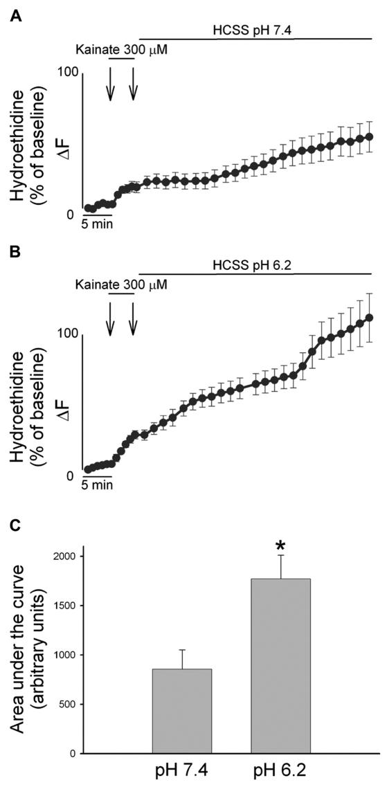
Figure 4.
AMPAR-mediated ROS generation is enhanced by low extracellular pH. A, B. Cortical cultures were loaded with HEt, and exposed for 5 min to 300 μM kainate and washed for 50 min with a buffer at pH 7.4 (A) or pH 6.2 (B). HEt fluorescence changes for each neuron are expressed as the ratio of fluorescence at each time point (Fx) to its own baseline fluorescence (F0). Traces show mean (± SEM) of 29 (A) or 40 (B) neurons from 3 experiments. C. Bar graph depicts the cumulative ROS production as area under the curve during the recovery phase in the two conditions, * indicates differences between washout at pH 7.4 versus washout at pH 6.2 (P < 0.01) Note the marked increase in HEt fluorescence only in the case of kainate exposures followed by an acidotic wash out.

