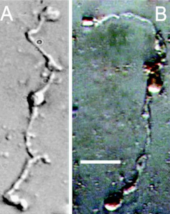
Figure 1.
DAB-labeled isolated rat Müller cells viewed using conventional light microscopy. (A) Control DAB-processed rat Müller cell in the absence of the ZnT-3 antibody showing no labeling. (B) ZnT-3-DAB-labeled Müller cell viewed using conventional light microscopy. Müller cell apical villi, soma, and endfeet show dark brown labeling indicating the presence of the ZnT-3 protein. Scale bar: 40 μm.

