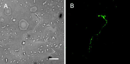
Figure 2.
FITC labeled ZnT-3 localization of mouse Müller cell viewed using confocal microscopy. (A) White light. (B) Fluorescent FITC. The ZnT-3 protein appears to be distributed ubiquitously in the FITC-labeled Müller cells. Slightly higher concentrations of fluorescent label appeared in endfoot and apical regions, while Müller cell somas typically labeled with lower intensities than other regions of the cell, as shown here. Scale bar: 40 μm.

