
Figure 1.
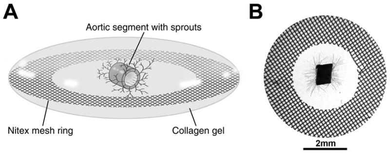
Depiction of the MRSG angiogenesis assay. A) Diagram showing an oblique view of an MRSG assembly comprised of a lenticular collagen gel, supportive Nitex mesh ring, and mouse aortic segment. B) View of an actual MRSG preparation after 7 days of culture. Microvascular sprouts are visible within the collagen gel.