
Figure 4.
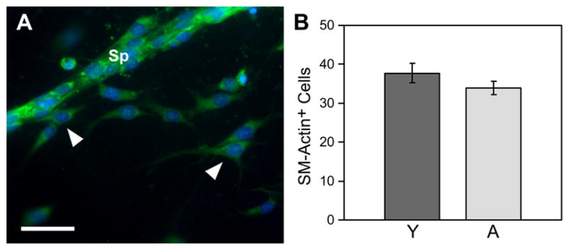
Outgrowth of SM actin-positive cells from young and aged aortic segments is similar. A) Sprout (Sp) from an aged aortic segment exhibits substantial numbers of SM actin-positive cells (green). Additionally, many SM actin-positive cells have migrated as single cells into the collagen gel (arrowheads). Cell nuclei are labeled with DAPI. The average number of SM actin-positive cells per 10X field that had invaded the collagen and were not in sprouts was similar in aortic segments from aged mice as that of segments from young mice, as represented quantitatively in panel B (vertical bars = standard error; p=>0.05, n = 4 for young and n = 5 for aged aortic segments). In A, scale bar = 100μm.