
Figure 1.
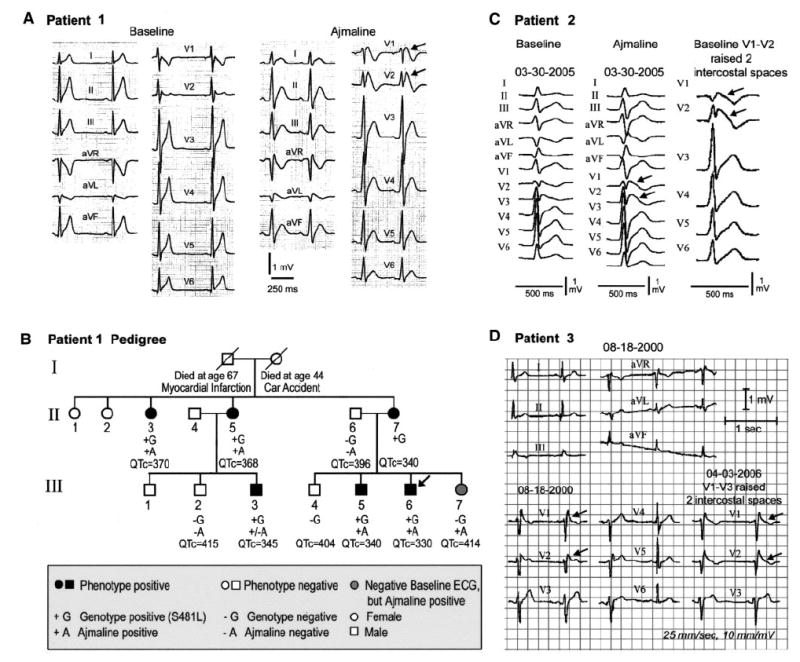
A, Twelve-lead ECG of patient 1 before and after ajmaline recorded with V1 and V2 displaced superiorly 2 intercostal spaces. B, Pedigree of family of patient 1 (III-6, arrow: proband). Arrows in ECGs depict prominent (type I) ST-segment elevation. C, Twelve-lead ECG of patient 2 before and after ajmaline (1 mg/kg) and at baseline with V1 and V2 displaced superiorly 2 intercostal spaces to unmask a type 1 ST-segment elevation. D, Twelve-lead ECG of patient 3 with V1 through V3 in normal position and displaced superiorly 2 intercostal spaces.