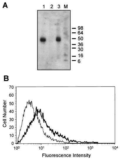
Figure 3.
Expression of recombinant A33 antigen by transfected Cos cells. Cos cells were transfected either with the parental vector, pcDNA3, or with pcDNA3 into which a 2.6-kb A33 antigen cDNA had been subcloned. Cells were harvested in this experiment 5 days after transfection and subjected to Western blot analysis (A) and flow cytometry (B). (A) Transfected cells were solubilized in Triton X-100 and electrophoresed into SDS/polyacrylamide gels without reduction and processed for Western blot analysis as described. The signal obtained with Cos cells transfected with pcDNA3 containing A33 antigen cDNA (lane 1) corresponds to approximately the same Mr (43,000) as the signal obtained with LIM1215 cells expressing high endogenous levels of A33 antigen (lane 3). Cells transfected with the parental vector gave no signal (lane 2). The positions of blue prestained standards (lane M) (NOVEX, San Diego) are indicated on the right. (B) FACScan profiles of control and A33 antigen-expressing Cos cells. The profile obtained with A33 antigen-expressing Cos cells (shown in bold) has been superimposed on the profile obtained with Cos cells transfected with pcDNA3 alone to show the shift to the right in fluorescence intensity of cells expressing A33 antigen.

