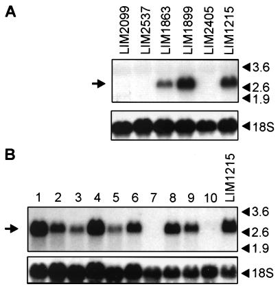
Figure 4.
Northern blot analysis of A33 antigen mRNA, indicated by arrows on the left, in (A) cell lines derived from human colorectal carcinoma and (B) normal and diseased human colonic tissue. (A) The six colorectal carcinoma cell lines had been previously analyzed for A33 antigen expression using immunocytochemistry and flow cytometry (unpublished data), and half (LIM1215, LIM1863, and LIM1899) were found to be positive. The remaining half (LIM2099, LIM2405, and LIM2537) were negative. The pattern of A33 antigen mRNA expression shown is consistent with the protein expression data. (B) A33 antigen mRNA expression in samples of normal and diseased human colorectal tissue obtained from patients during surgical resection. The samples are in pairs except for lanes 1–3, which contain RNA extracted from the normal colon, adenomatous polyp, and tumor of the same patient, respectively. Thus, lanes 1, 4, 6, and 8 contain RNA extracted from the crypts of normal colonic mucosa, and the strong hybridization signals correspond to relatively high expression of A33 antigen compared with that in the corresponding tumor preparations (lanes 3, 5, and 7) and the RNA extracted from the large polyp (lane 2), which all produced weaker A33 antigen mRNA signals. Lane 9 contains RNA extracted from the inflamed colonic crypts of a patient with Crohn disease, which produces an A33 antigen signal similar to that of its normal counterpart (lane 8). Lane 10 contains RNA from human peripheral blood buffy coat cells, and lane 11, RNA from LIM1215 cells (positive control). For both blots, the lower panel shows the pattern obtained with a γ-32P-labeled oligonucleotide probe designed to hybridize to 18 S rRNA. Arrows on the right indicate the positions of RNA markers.

