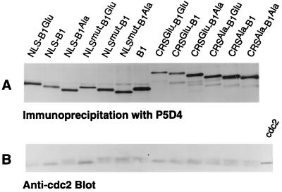
Figure 2.
Expression and cdc2 binding of cyclin B1 fusion proteins. Microinjected oocytes were labeled for 5 h as described (13) using 0.5 mCi/ml (1 Ci = 37 GBq) [35S]Met and 0.25 mCi/ml [35S]Cys. (A) Oocytes were lysed and half of the lysate was subjected to immunoprecipitation to recover labeled cyclin proteins using a mAb directed against the epitope tag. Proteins were analyzed by 12.5% SDS/PAGE and detected by fluorography. (B) The other half of each sample was immunoprecipitated as in A, and cdc2 proteins were detected by immunoblotting as described. The last lane in B shows in vitro translated cdc2 as a control.

