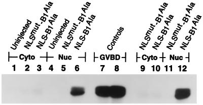
Figure 4.
Localization of NLS–B1Ala and NLSmut–B1Ala associated H1 kinase activity. Nuclei were manually isolated at either 2.5 or 4.5 h from oocytes microinjected with in vitro-synthesized RNA encoding NLS–B1Ala and NLSmut–B1Ala. Nuclear and cytoplasmic fractions were then immunoprecipitated using P5D4 antibody to recover the epitope-tagged cyclin B1 fusion proteins, and immunoprecipitates were examined for histone H1 kinase activity, associated with active MPF. Phosphorylated histone H1 was detected by autoradiography after separation by 15% SDS/PAGE. Samples from nuclei (Nuc) or from enucleated oocytes (Cyto) were analyzed at two different time points after microinjection; 2.5 h (lanes 2, 3, 5, and 6) and 4.5 h (lanes 9–12). As a negative control, the cytoplasmic (lane 1) and nuclear (lane 4) fractions from uninjected oocytes were similarly analyzed. For lanes 1–6 and 9–12, each sample corresponds to three nuclei or enucleated oocytes. As a positive control, oocytes that reached GVBD (at ≈5.5 h) after microinjection with RNA encoding NLS–B1Ala were similarly analyzed for histone H1 kinase activity. Lanes 7 and 8 correspond to one-quarter and to one-half of an oocyte, respectively.

