Figure 2.
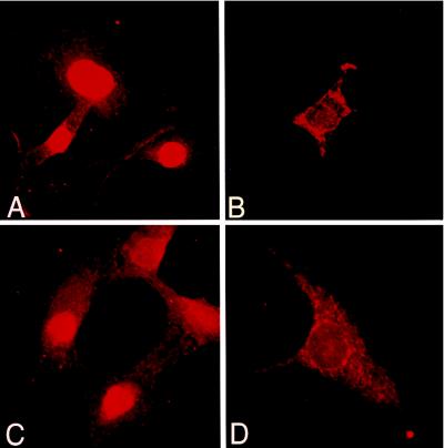
Differential distributions of N- and C-terminal IL-1α segments in stably transfected MC. (A) Intense nuclear staining in cells transfected with the 16-kDa N-terminal IL-1α propiece, stained with N-terminal-specific antibody. (B) Transfected cells expressing K82K83 → Thr82Thr83 mutated N-terminal IL-1α propiece reveal intense cytosolic staining, with minimal nuclear staining using N-terminal-specific antibody. (C) Transfected cells expressing the 31- to 33-kDa IL-1α precursor exhibit bright nuclear staining with the N-terminal-specific antibody; with the C-terminal-specific antibody (D), staining is limited to the cytosol. (A–D; ×750.)

