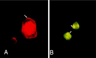
Figure 3.
Differential distribution of the N- and C-terminal segments of the IL-1α precursor in phorbol ester-induced HL-60 human promyelocytic leukemia cells. (A) Induced cells exhibit intense nuclear staining, as well as plasma membrane staining (white arrow) when probed with the N-terminal-specific antibody. (B) The C-terminal-specific antibody yields only cytosolic staining (arrows). (A, ×700; B, ×270.)

