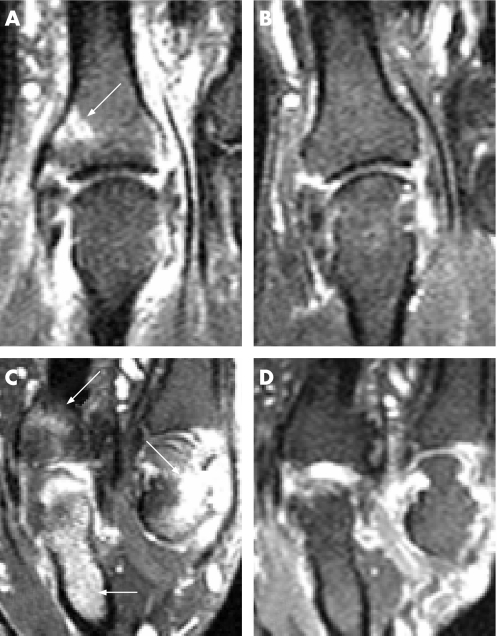
Figure 1 Coronal T1‐weighted post‐gadolinium‐diethylenetriamine penta‐acetic acid fat‐suppressed images of the second (A,B) and fourth–fifth (C,D) metacarpophalangeal (MCP) joints from two of the study patients pre–and‐post treatment with infliximab. (A) The white arrow shows entheseal bone oedema distal to the joint space and there is high signal around the second MCP joint representing active synovitis‐both of which is resolved in the post‐treatment scan (B). (C) Pretreatment scan of the second patient. There is high signal representing extensive bone oedema in the proximal phalanx of the fourth finger. There is also bone oedema distal to the joint space on the fourth MCP compatible with an entheseal location, and on the proximal fifth MCP joint there is an erosion with bone oedema (white arrows). These lesions have resolved in the post‐treatment scan (D).

An official website of the United States government
Here's how you know
Official websites use .gov
A
.gov website belongs to an official
government organization in the United States.
Secure .gov websites use HTTPS
A lock (
) or https:// means you've safely
connected to the .gov website. Share sensitive
information only on official, secure websites.
