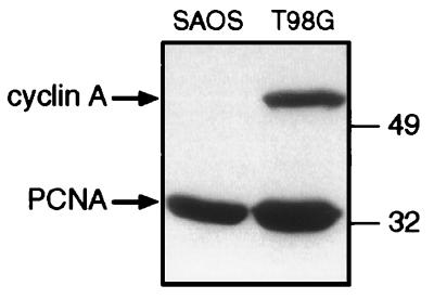
Figure 4.
Cyclin A protein levels in SAOS-2 and T98G cell lines. Nuclear extracts were prepared and 50 μg of protein were loaded onto a 10% acrylamide/SDS minigel. After blotting onto nitrocellulose the membrane was incubated at the same time with an antibody against cyclin A (Oncogene Science) and one against proliferating cell nuclear antigen (PCNA) (Santa Cruz).

