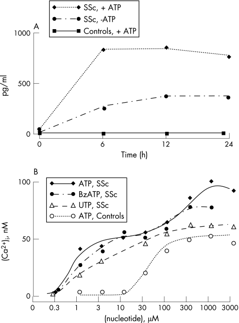
Figure 1 Fibroblasts from patients with SSc are hypersensitive to stimulation with extracellular ATP. (A) Fibroblasts from healthy controls and patients with SSc were placed in 24‐well plates at a concentration of 4×10⁵/ml in a volume of 0.5 ml. ATP was added at a concentration of 1 mmol/l. At the end of the incubation, supernatants were assayed for IL‐6 content by ELISA. (B) Fibroblasts were layered onto glass coverslips, loaded with Fura‐2/AM and transferred to a fluorimeter cuvette. After 5 minutes, nucleotides were added and peak [Ca2+]i over basal measured. For other experimental details see Solini et al.4

An official website of the United States government
Here's how you know
Official websites use .gov
A
.gov website belongs to an official
government organization in the United States.
Secure .gov websites use HTTPS
A lock (
) or https:// means you've safely
connected to the .gov website. Share sensitive
information only on official, secure websites.
