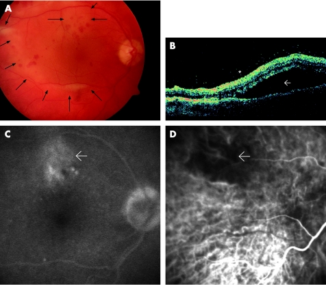
Figure 1 Case 1. Left eye:(A) Fundus photograph showing chorioretinitis with placoid, pale, yellowish lesions, subretinal fluid accumulation (→) and intraretinal haemorrhages. (B) Optical coherence tomography revealing extensive serous detachment of the posterior pole (→) and condensed vitreous close to the retinal surface (*) (C) Late‐phase fluorescein angiography disclosed late staining at the level of the retinal pigment epithelium that was most prominent in the areas of yellowing. (D) Indocyanine green angiography showing early hypofluorescence in the same area (→).

An official website of the United States government
Here's how you know
Official websites use .gov
A
.gov website belongs to an official
government organization in the United States.
Secure .gov websites use HTTPS
A lock (
) or https:// means you've safely
connected to the .gov website. Share sensitive
information only on official, secure websites.
