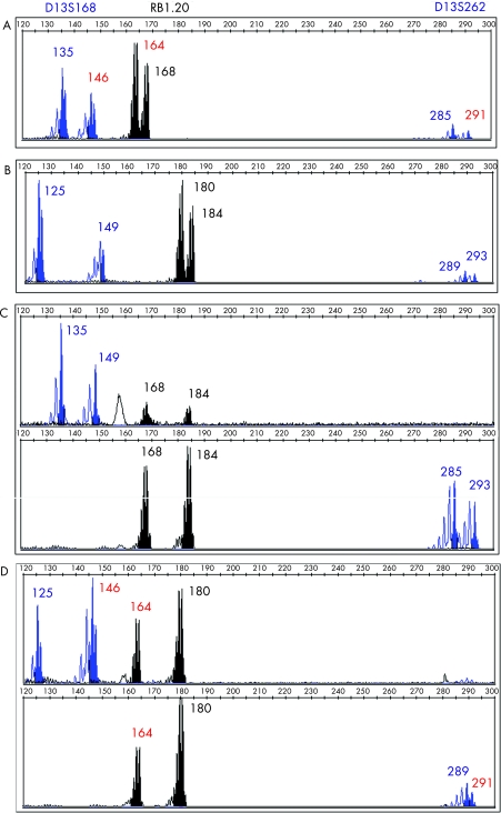
Figure 2 Electrophoretograms from the DNA of proband (A), her partner (B), blastomeres from E8, normal embryo (C) and E6, embryo carrying the mutation (D) amplified with three markers: D13S168, RB1.20 and D13S262. Allelic length sizes are given in base pairs. The allelic sizes in red correspond to the maternal chromosome carrying the mutation.

An official website of the United States government
Here's how you know
Official websites use .gov
A
.gov website belongs to an official
government organization in the United States.
Secure .gov websites use HTTPS
A lock (
) or https:// means you've safely
connected to the .gov website. Share sensitive
information only on official, secure websites.
