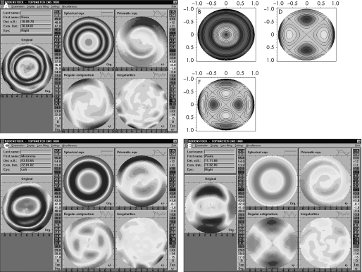
Figure 5 Decomposition of corneal topography (by Fourier analysis) into the four main aberration components (spherical‐like, coma‐like, regular astigmatism and irregular astigmatism) for a spherical (A), an elliptical (B) and a mixed ablation (C). Note the similarity between the regular astigmatic component and the pure secondary regular astigmatism (horizontal) representations in the Zernike pyramid on the right side for the elliptical ablation (sixth order horizontal astigmatism, B) and mixed ablation (fourth order horizontal astigmatism, C).

An official website of the United States government
Here's how you know
Official websites use .gov
A
.gov website belongs to an official
government organization in the United States.
Secure .gov websites use HTTPS
A lock (
) or https:// means you've safely
connected to the .gov website. Share sensitive
information only on official, secure websites.
