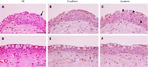
Figure 2 H&E staining (A, D) and immunodetection of E‐cadherin (B, E) and β‐catenin (C, F) in the human pterygium head (A–C) and body (D–F). In the pterygium head, thickness is relatively marked compared with the body (A, D). The head epithelium consists of squamous metaplasia with several goblet cells (A, arrow) in the superficial layer. E‐cadherin (B) as well as β‐catenin (C) is predominantly expressed in a variety of epithelial cells with circumferential cytoplasmic and apical membrane patterns. Several epithelial cells show dense nuclear and cytoplasmic immunoreactivity for β‐catenin (C, arrowheads). In the pterygium body, the epithelium consists of a few layers with round cells including goblet cells (D). The thickness was relatively thin compared to the head. Immunoreactivity for E‐cadherin (E) and β‐catenin (F) is detected in the epithelium, but not in the stroma, with an apical membrane pattern. Bar = 50 μm.

An official website of the United States government
Here's how you know
Official websites use .gov
A
.gov website belongs to an official
government organization in the United States.
Secure .gov websites use HTTPS
A lock (
) or https:// means you've safely
connected to the .gov website. Share sensitive
information only on official, secure websites.
