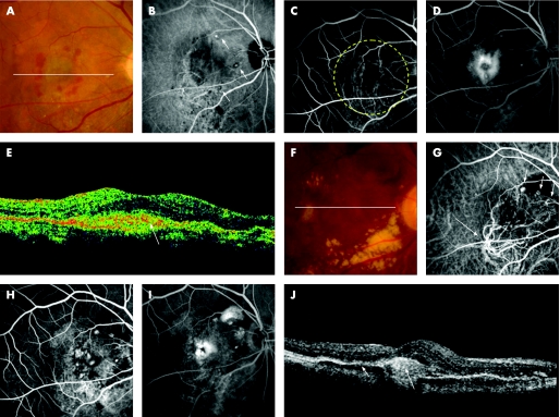
Figure 2 A 63‐year‐old woman (patient 8) was referred to our clinic with a six‐month history of metamorphopsia and decreased visual acuity in the left eye (OS). At the initial visit, her visual acuity was 20/30 OS and funduscopic examination revealed a reddish‐orange nodule with overlying greyish material in the posterior pole (A). Indocyanine green angiography (IA) reveals a branching vascular network terminating in polypoidal lesions, which are juxtafoveal in location (B). Fluorescein angiography (FA) shows classic juxtafoveal choroidal neovascularisation (CNV) corresponding to the polypoidal lesion seen on IA (C, early phase; D, late phase). A sectional image with optical coherence tomography (OCT) along the white line shows a polypoidal lesion (arrow) and adjacent subretinal material with moderate reflectivity (long arrow) (E). She was treated with photodynamic therapy (PDT) in the left eye. The yellow dotted line indicates the laser irradiation spot. A fundus photograph at three months after PDT shows neither reddish‐orange nodules nor overlying greyish material (F). IA shows no polypoidal lesions (G). FA shows no classic CNV (H, early phase; I, late phase). OCT image along the white line shows reduced polypoidal lesion (arrow) (J). Her visual acuity was 20/13 OS.

An official website of the United States government
Here's how you know
Official websites use .gov
A
.gov website belongs to an official
government organization in the United States.
Secure .gov websites use HTTPS
A lock (
) or https:// means you've safely
connected to the .gov website. Share sensitive
information only on official, secure websites.
