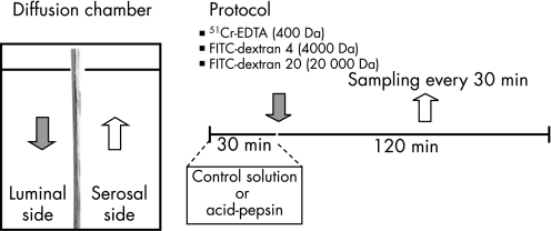
Figure 1 Schematic representation of permeability studies and protocol. Oesophageal mucosal sections of approximately 0.5 cm2 were cut and mounted in a diffusion chamber. The diffusion chamber allowed for exposure of the luminal side of the tissue to different test solutions and regular sampling from the serosal side to detect the degree of mucosal permeability to different molecules.

An official website of the United States government
Here's how you know
Official websites use .gov
A
.gov website belongs to an official
government organization in the United States.
Secure .gov websites use HTTPS
A lock (
) or https:// means you've safely
connected to the .gov website. Share sensitive
information only on official, secure websites.
