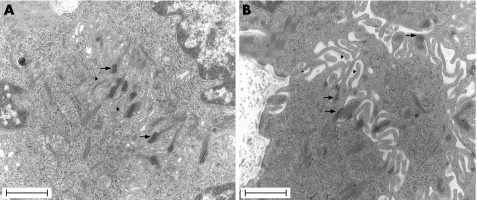
Figure 7 Transmission electron microscopy micrographs of rat oesophageal epithelium in (A) control rat and (B) stressed rat. An important loss of contact between cells only maintained in the desmosome region (arrows) was observed in stressed rats (B). Microvillous processes (mark) . Scale bars = 1 μm.

An official website of the United States government
Here's how you know
Official websites use .gov
A
.gov website belongs to an official
government organization in the United States.
Secure .gov websites use HTTPS
A lock (
) or https:// means you've safely
connected to the .gov website. Share sensitive
information only on official, secure websites.
