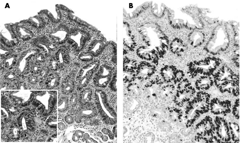
Figure 2 (A–B) Low‐grade non‐invasive neoplasia (NiN): H&E stain (A) shows all the spectrum of the cytohistological lesions of the NiN (altered architecture, atypical/pseudostratified nuclei, loss of superficial differentiation). (B) Mib1 nuclear immunostain is clearly demonstrated throughout the glandular units (from the superficial to the “basal” epithelia; original magnification: ×40).

An official website of the United States government
Here's how you know
Official websites use .gov
A
.gov website belongs to an official
government organization in the United States.
Secure .gov websites use HTTPS
A lock (
) or https:// means you've safely
connected to the .gov website. Share sensitive
information only on official, secure websites.
