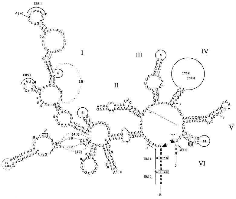
Figure 2.
Group II intron secondary structure of the Asplenium nidus nad2 intron with the exchanges in Marsilea drummondii given next to the model, in parentheses, and with dotted lines, respectively. Roman numerals indicate the six well-defined domains radiating from a central wheel (8). Splice sites are indicated by arrows. Tertiary structure interactions between the exon and intron binding sites (EBS1–IBS1, EBS2–IBS2) and other nucleotides (γ–γ′, δ–δ′) are highlighted. A guanosine residue (encircled) is found at the branch-site position where adenosine is conserved in other introns. Editing is required at the 3′ end of the upstream exon to reconstitute conserved codons and to allow the EBS–IBS interactions.

