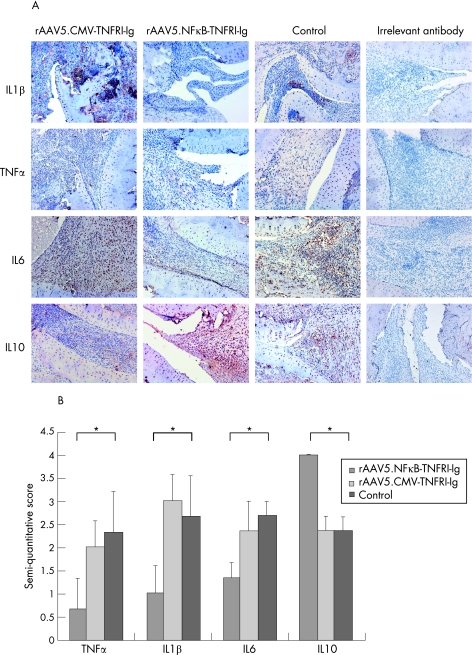
Figure 4 Cytokine levels in rat synovial tissue. At the end of the experiment, ankle joints were collected, embedded in paraffin wax and immunohistochemically stained with anti‐rat interleukin (IL)1β IL6 and IL10 and tumour necrosis factor (TNF)α, antibodies and counterstained with Mayer's haemalum. An irrelevant antibody was used as control. (A) Representative pictures are shown (original magnification 100×). (B) The images were semiquantitatively scored (0–4) by two independent observers (*p<0.05). Values are expressed as mean (SEM) (n = 5/group).

An official website of the United States government
Here's how you know
Official websites use .gov
A
.gov website belongs to an official
government organization in the United States.
Secure .gov websites use HTTPS
A lock (
) or https:// means you've safely
connected to the .gov website. Share sensitive
information only on official, secure websites.
