Figure 1.

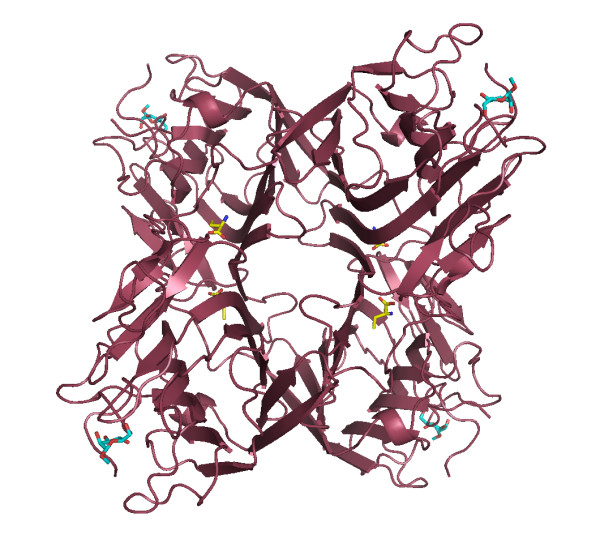
Crystal structure of CGL-αMM. The vision of the Abu position can be seen in the canonical dimmer between the monomers at hydrophobic cavity. Abu is displayed in yellow and α-methyl-mannoside is in blue.

Crystal structure of CGL-αMM. The vision of the Abu position can be seen in the canonical dimmer between the monomers at hydrophobic cavity. Abu is displayed in yellow and α-methyl-mannoside is in blue.