
Figure 2. The pCaSpeR5 and pUAS-C5 vectors are functional.
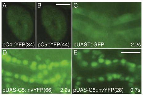
(A and B) Constructs expressing a yellow fluorescent protein (YFP) (3) driven by the glass multimer reporter (GMR) enhancer element (5) produce comparable expression in adult eyes of transgenic animals made using (A) pCaSpeR4 (pC4::YFP) and (B) pCaSpeR5 (pC5::YFP) vectors. Representative lines are shown with line numbers in parenthesis. Exposure times for A and B were 2.2 s and were made using a Model MZFLIII microscope (Leica Microsystems GmbH, Wetzlar, Germany), a Model C4742-95 camera (Hamamatsu Photonics K.K, Hamamatsu, Japan), and a green fluorescent protein (GFP) filter set (Chroma Technology, Rockingham, VT, USA). Scale bar for A and B in panel B, 25 μm. (C E) A nuclear-localized venus-YFP (nvYFP) (3) construct in pUAS-C5 driven by the tracheal-specific btl-Gal4 appears brighter (D and E) than the widely used pUAST-GFP driven by btl-Gal4 (C) (4). Images are of live stage 16 embryos heterozygous for each of the driver and responder constructs. Images were captured using an AxioPlan II microscope, an AxioCam camera (both from Carl Zeiss, Jena, Germany), and a GFP filter set using the indicated exposure times. Scale bar for C E in panel E, 5 μm.