
Fig. 4. MBP-primed T lymphocytes induce the expression of iNOS in mouse BV-2 microglial cells.
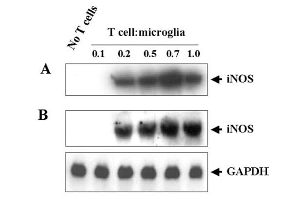
BV-2 cells were stimulated with different concentrations of MBP-primed T cells under serum-free conditions. In A, cell homogenates were electrophoresed, transferred on nitrocellulose membrane, and immunoblotted with antibodies against mouse macrophage iNOS as described under “Materials and Methods.” In B, after 6 h of incubation, Northern blot analysis for iNOS mRNA was carried out as described under “Materials and Methods.” GAPDH, glyceraldehyde 3-phosphate dehydrogenase.