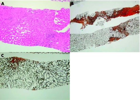
Figure 2 (A) Biopsy specimen showing marked sinusoidal dilatation, hepatocyte atrophy and an area of scarring with linking of two portal tracts and no visible hepatic vein (H&E stain, ×200 magnification). (B) There is a diffuse pattern of sinusoidal fibrosis, which is irregularly distributed throughout the acinus. The cell plate pattern is deranged with atrophic and twinned cell plates in close proximity (Gordon and Sweet reticulin stain, ×125 magnification). (C) Broad scars of doubly refractile collagen (red–brown) distorting the architecture and causing cirrhosis (Gordon and Sweet reticulin stain, ×125 magnification).

An official website of the United States government
Here's how you know
Official websites use .gov
A
.gov website belongs to an official
government organization in the United States.
Secure .gov websites use HTTPS
A lock (
) or https:// means you've safely
connected to the .gov website. Share sensitive
information only on official, secure websites.
