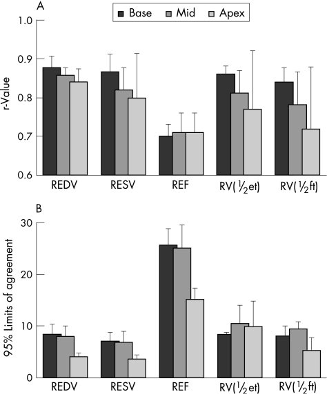
Figure 3 Comparisons between the real‐time three‐dimensional echocardiography derived indices of regional left ventricular function against cardiac magnetic resonance (CMR) reference values calculated for basal, mid and apical segments: (A) Pearson's correlation coefficients and (B) limits of agreement. Bars represent segmental data averaged over the 31 study patients. REDV, regional end‐diastolic volumes; REF, regional ejection fraction; RESV, regional end‐systolic volumes; RV(½et), regional volume at half ejection; RV(½ft), regional volume at half filling times.

An official website of the United States government
Here's how you know
Official websites use .gov
A
.gov website belongs to an official
government organization in the United States.
Secure .gov websites use HTTPS
A lock (
) or https:// means you've safely
connected to the .gov website. Share sensitive
information only on official, secure websites.
