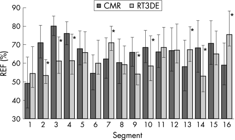
Figure 4 Cardiac magnetic resonance (CMR) derived and real‐time three‐dimensional echocardiography (RT3DE) derived mean regional ejection fraction (REF) values in 16 segments obtained in 15 patients with normal wall motion. The upward error bars represent the SD, whereas the tips of the downward error bars point at the optimised REF threshold values for each imaging modality (*p<0.05 vs CMR).

An official website of the United States government
Here's how you know
Official websites use .gov
A
.gov website belongs to an official
government organization in the United States.
Secure .gov websites use HTTPS
A lock (
) or https:// means you've safely
connected to the .gov website. Share sensitive
information only on official, secure websites.
