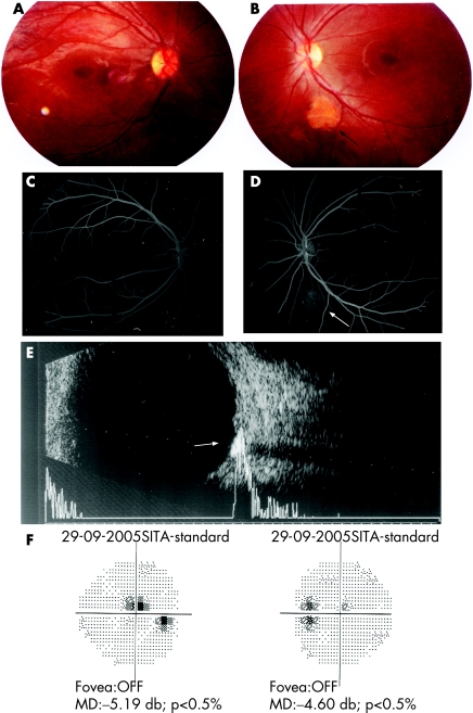
Figure 1 (A,B) Fundus photographs of right and left eyes revealing bilateral bull's eye maculopathy and left juxta papillary calcific lesion (arrow). (C,D) Fundus fluorescein angiography of right and left eyes. Bilateral dark choroids and hyperfluorescent juxtapapillary lesion in left eye. (E) B ultrasound scan reveals shadowing posterior to the highly dense lesion (A ultrasound scan: sound attenuation posterior to the lesion). (F) Perimetric central field defects. MD, mean deviation.

An official website of the United States government
Here's how you know
Official websites use .gov
A
.gov website belongs to an official
government organization in the United States.
Secure .gov websites use HTTPS
A lock (
) or https:// means you've safely
connected to the .gov website. Share sensitive
information only on official, secure websites.
