
Figure 7.
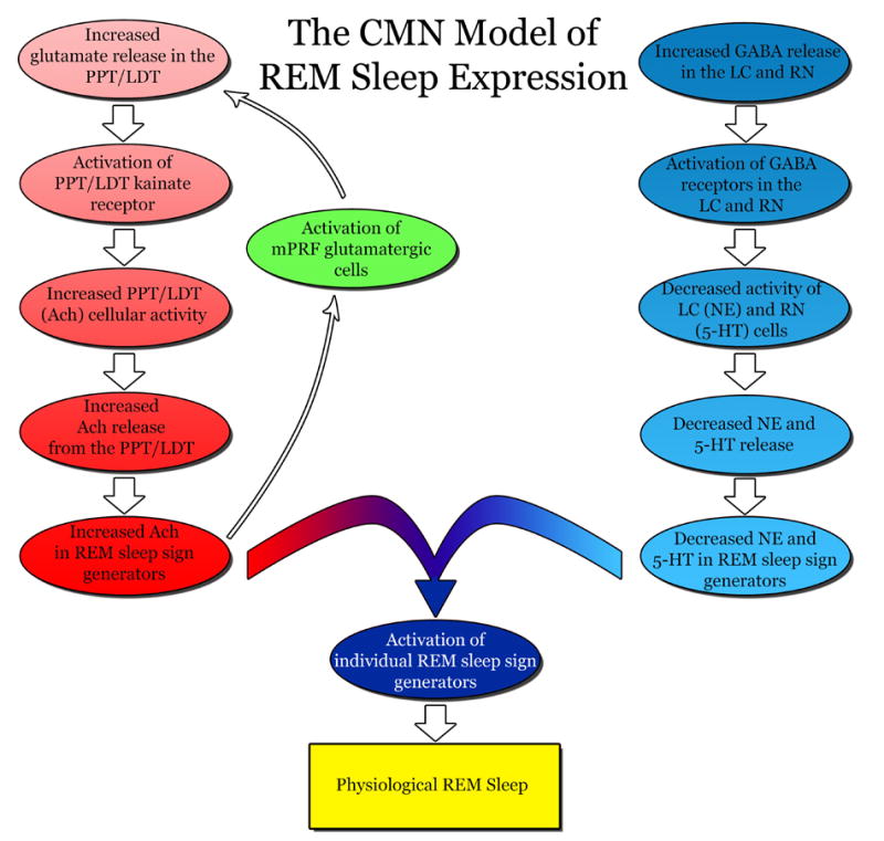
Cellular-Molecular-Network model of cholinergic and aminergic activity for the expression of physiological REM sleep. For the activation of an individual REM sleep sign generator, an increase in acetylcholine (Ach) is accompanied by a decrease in norepinephrine (NE) and serotonin (5-HT) within each generator. An increase in Ach (red) is the result of increased glutamate release in the pedunculopontine tegmentum (PPT) and the lateral dorsal tegmentum (LDT). This increased glutamate activates kainate receptors on the PPT/LDT and signals the release of Ach. The increased Ach in the sleep sign generators also activate medial pontine reticular formation (mPRF) cells that release glutamate (green) to the PPT/LDT and facilitate the continued release of Ach during REM sleep (maintenance of REM sleep episode). A decrease in NE and 5-HT (blue) is the result of increased γ-aminobutyric acid (GABA) release in the locus coeruleus (LC) and raphe nucleus (RN), respectively. The increased GABA reduces the presence of NE and 5-HT by inhibiting the cellular activity of the LC and RN. The combination of decreased NE and 5-HT and increased Ach in each individual REM sleep sign generator ultimately generates the expression of physiological REM sleep.