
Figure 2. Electron microscopic images of left ventricle and papillary muscles from control (A, B) and AMPKα2−/− (C–F) mice.
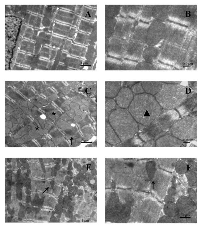
(A) Overview of a control myocyte in longitudinal section, with mitochondria and myofibrils arranged in regular longitudinal columns. (C and E) Longitudinal section of cardiomyocytes from papillary (C) and ventricular (E) muscles from AMPKα2−/− mice showing myofibrillar disorganization, and irregular arrangement of intermyofibrillar mitochondria with clusters of mitochondria of variable size. (B) Detail of sarcomeres in a control myocyte, showing mitochondria tightly packed along sarcomeres. (D and F) Details of sarcomeres in AMPKα2−/− myocytes from papillary (D) and ventricular (F) muscles, showing dense packing of mitochondria of irregular size. Asterisk: large mitochondria having irregular shape. Arrows: splitting of myofibrils. Arrowhead: dividing mitochondrion.