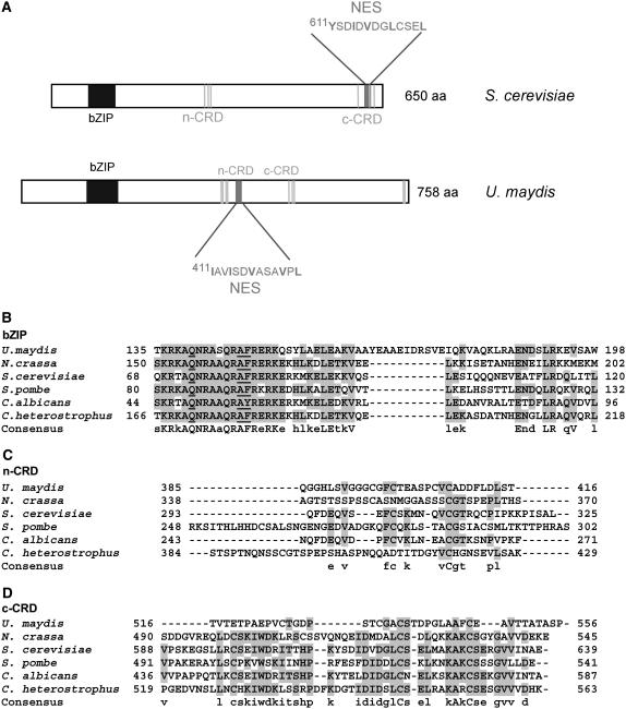
Figure 1.
Domain Organization of Yap1.
(A) Yap1p of S. cerevisiae and Yap1 of U. maydis are compared. The vertical gray lines indicate the position of Cys residues. NES (dark-gray bar), nuclear export sequence; aa, amino acids.
(B) Alignment of the bZIP domain of AP-1–like proteins from U. maydis, Neurospora crassa, S .cerevisiae, S. pombe, C. albicans, and C. heterostrophus. Accession numbers for these proteins are in Methods. Uppercase letters indicate identity in all proteins compared, and lowercase letters indicate that three or more proteins carry this residue.
(C) Alignment of the n-CRD domains of proteins listed in (A). Shading follows the scheme given in (A).
(D) Alignment of the c-CRD domains of proteins listed in (A). Numbers give amino acid positions in the respective proteins.

