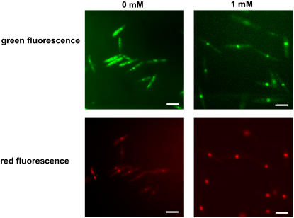
Figure 4.
Subcellular Localization of Yap1 in the Presence of H2O2.
Strain FB2yap1:3XeGFPcbx:pANNE3090 was cultured in liquid CM-glucose medium, exposed to the indicated concentrations of H2O2 for 1 h, and assayed microscopically for GFP and RFP fluorescence. FB1yap1:3XeGFPcbx:pANNE3090 displayed the same behavior (data not shown). Bars = 10 μm.

