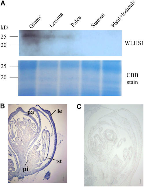
Figure 8.
Protein Gel Blotting Analysis and Distribution of WLHS1 Proteins in Wheat Inflorescences.
(A) Protein gel blotting of individual parts of floral organs at the prebooting stage. Total proteins were extracted from each tissue and separated by SDS-PAGE. The proteins were stained either with Coomassie blue or, after membrane transfer, with antibodies raised against the C-terminal peptide of WLHS1. The antibody produced a band at ∼26 kD, corresponding to the WLHS1-B and/or WLHS1-D protein. No bands were seen at 19 kD, which corresponds to the expected size of the WLHS1-A protein.
(B) Immunolocalization of WLHS1. Strong staining is seen in the palea and lemma. Bar = 100 μm. pi, pistil; st, stamen; pa, palea; le, lemma.
(C) Control section. No staining was seen using serum from a preimmunized rabbit.
Bar = 100 μm.

