Figure 5.
Gravity-Induced Auxin Asymmetry in Wild-Type and mdr4 Roots.
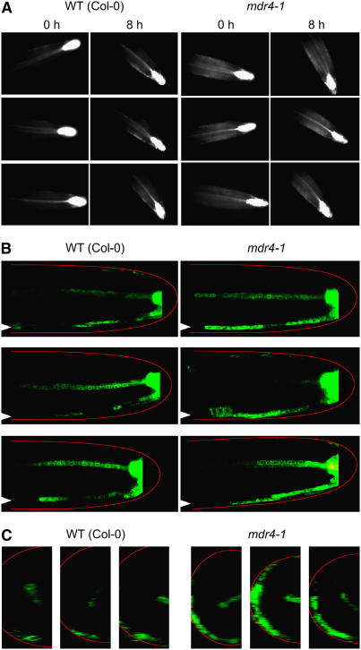
(A) ProDR5:GFP signal in wild-type and mdr4 roots before and after 8 h of reorientation. Images were obtained with a horizontally mounted epifluorescence microscope. Three example experiments are shown. The background auxin-dependent GFP signal in the elongation zone was generally higher in mdr4 roots, and the accumulation on the lower flank after gravitropism was more diffuse than in the wild type.
(B) Optical slices (longitudinal medial) through wild-type or mdr4 root apices expressing ProDR5:GFP obtained by laser scanning confocal microscopy after 6 h of gravitropism. A generalized root outline in red is superimposed for orientation because the intense ProDR5:GFP signal landmark at the root tip is omitted. Arrowheads at the left edge of each image point to the ProDR5:GFP signal along the lower flank of the root, which differs between the mutant and the wild type. Shown are three individuals that are representative of the six examined for each genotype.
(C) Cross sections computationally constructed from the z-series of confocal images from which the images in (B) were selected show greater ProDR5:GFP signal in the lower part of mdr4 roots compared with the wild type.

