
Figure 5.
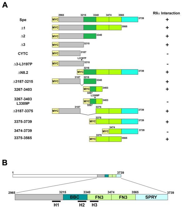
A, Schematic diagram of select Myc-tagged Myospryn constructs expressed in COS cells to map the RIIα-binding domain. Interactions results are shown at right (+, positive interaction; -, negative interaction). B, Schematic depiction of the three PKA anchoring motifs in relation to the TRIM region of Myospryn. The H1 helix is localized immediately upstream of the TRIM region; H2 is found within the B-box coiled coil (BBC) domain; and H3 is situated within the first fibronectin 3 repeat (FN3).