
Figure 2.
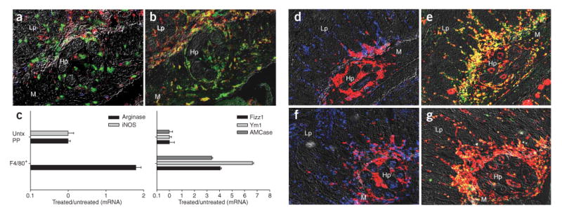
AAMac accumulation 4 d after H. polygyrus challenge is Stat6 dependent. (a,b) Four days after primary infection, Gr-1+ neutrophils (green) accumulated adjacent to the parasite, with substantially fewer CD4+ T cells (blue), IL-4Rhi cells (red), F4/80+ macrophages (b, red) and CD206+ cells (green) compared to what is seen after secondary infection (see Fig. 1d). (c) F4/80+ cells dissected from the host-parasite interface by IF-LCM 4 d after challenge showed gene expression profiles characteristic of AAMacs, including undetectable iNOS (left) and high levels of arginase, Fizz1 (right), Ym1 and AMCase. Mean and s.e.m. of four or five mice per treatment group is plotted on log scales. (d–g) CD4+ T cells transferred from H. polygyrus–primed mice induced alternatively activated macrophages in wild-type (d,e) but not Stat6-/- recipients (f,g). Gr-1+ neutrophils (red) and CD4+ T cells (blue) accumulated around the parasite 4 d after infection following adoptive transfer of TH2 memory cells into wild-type (d) and Stat6-/- (f) recipients. Serial tissue sections show staining for macrophages (F4/80, red) and macrophage mannose receptor (CD206, green), showing that wild-type recipients (e) developed alternatively activated macrophages (yellow), whereas Stat6-/- recipients (g) did not. Lp, lamina propria; m, muscularis; Hp, H. polygyrus.