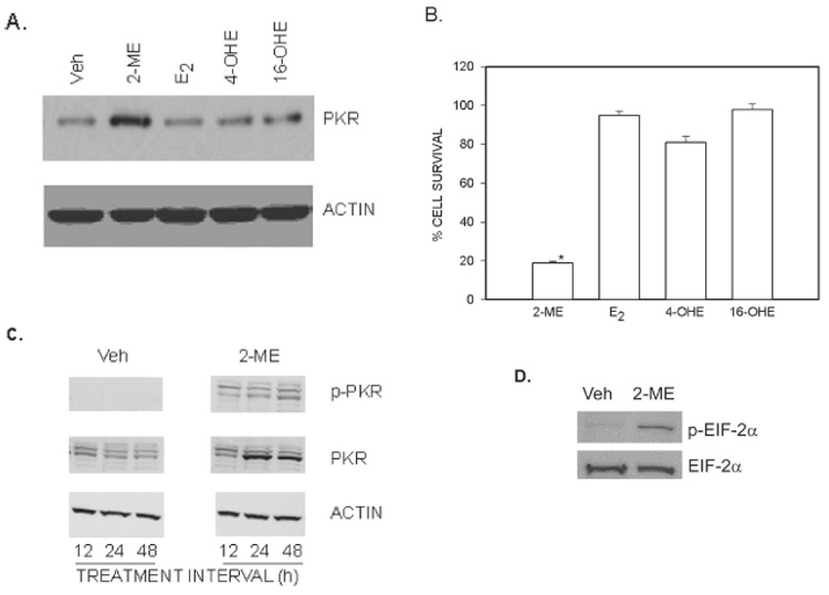
FIG. 1.

Association between an increase of PKR protein expression and cell survival in MG63 osteosarcoma cells. (A) PKR expression. Cytoplasmic extracts prepared from cells treated for 24 h with vehicle (Veh) or 10 µM of 2-ME, E2, 4-OHE, and 16-OHE was analyzed by Western blot hybridization using anti-PKR antibody and anti-actin antibody (Sigma). (B) Cell survival. Cells were treated with Veh or 5 µM of 2-ME, E2, 4-OHE, and 16-OHE for 72 h. The cells were harvested, and the viable cell counts were taken after staining with trypan blue. Values are mean ± SE (N = 3 replicate cultures). *p ≤ 0.05 vs. Veh. (C) Time-course of 2-ME effects on PKR. Cytoplasmic extracts prepared from cells treated for 12, 24, and 48 h with Veh or 10 µM of 2-ME were analyzed by Western blot hybridization using anti-phospho-PKR (pT446; Epitomics), anti-PKR (Santa Cruz Biotechnology), and anti-Actin (Sigma) antibodies. (D) EIF-2α phosphorylation. Cytoplasmic extracts prepared from cells treated with Veh or 10 µM of 2-ME for 24 h were analyzed by Western blot hybridization using anit-phospho-eIF-2α (pT446; Epitomics) and anti-eIF-2α (Cell Signaling Technology).
