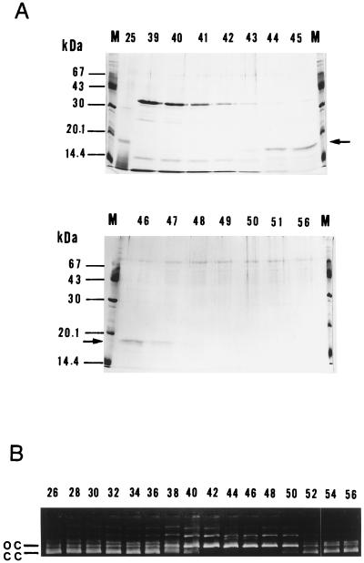
Figure 1.
Purification of UV endonuclease. M. luteus ATCC4698 cells (≈80 g wet weight) were fractionated as described, and the final DNA cellulose step was monitored for protein profiles and enzyme activity. (A) SDS/PAGE. Aliquots (40 μl) of fractions were analyzed on 12.5% gels, followed by staining with Coomassie blue. Figures, fraction numbers; arrows, the 1.8-kDa band; M, molecular mass standards. (B) UV endonuclease activity. Aliquots (3 μl) of fractions were subjected to UV endonuclease assay using the pUC18 substrate irradiated with 254-nm UV. Figures, fraction numbers; cc, closed circles; oc, open circles.

