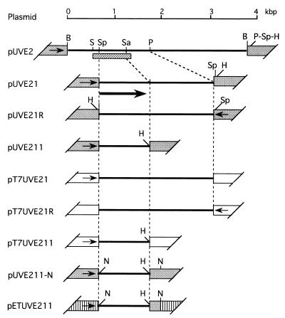
Figure 2.
Plasmid constructs. pUVE2 (see text) was digested with PstI and self-ligated. The resulting plasmid was cut with SphI, and the 1.1-kbp fragment formed was recloned into pUC18 to give pUVE21 and its reverse-oriented partner pUVE21R. pUVE21 was digested with SacII and HindIII, and the resulting larger fragment was blunt-ended and circularized with a phosphorylated HindIII linker (Takara, Kyoto) to give pUVE211. For the pT7T3 18U-based constructs, the SphI–HindIII fragments were prepared from pUVE21, pUVE21R, and pUVE211, and ligated to pT7T3 18U linearized with SphI and HindIII to generate pT7UVE21, pT7UVE21R, and pT7UVE211, respectively. For construction of pETUVE211, the SphI site of pUVE211 was converted to an NdeI site to make pUVE211-N, and its smaller NdeI fragment was inserted into the NdeI site of pET11a. Solid lines, M. luteus sequences; grey trapezoids, pUC18 sequences (arrows, orientation of lac promoter); open trapezoids, pT7T3 18U sequences (arrows, orientation of T7 promoter); hatched trapezoids, pET11a sequences (arrow, orientation of T7 promoter); hatched bar, sequenced region; thick arrow, the open reading frame identified. Restriction sites: B, BamHI; H, HindIII; N, NdeI; P, PstI; S, SalI; Sa, SacII; Sp, SphI.

