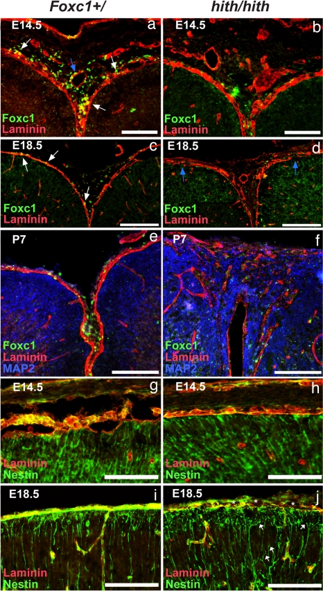
Fig. 5.
Progressive breakdown of midline BM and development of cortical dysplasia in Foxc1hith/hith animals. (a and b) Laminin (red) and Foxc1 (green) expression at the cortical midline in Foxc1+/ tissue at E14.5 shows Foxc1+ cells closely associated with the laminin BM (white arrows) and around laminin+ blood vessels (blue arrow). At this early age, the laminin BM is intact in Foxc1hith/hith brains and weakly Foxc1+ cells are associated with both the meninges and blood vessels at the midline. (c and d) At E18.5, a continuous BM surrounds the cortical tissue and Foxc1+ cells are embedded within the pial BM (white arrows). In the Foxc1hith/hith brain, breeches in the laminin BM are apparent at E18.5 (blue arrows), and, although some weakly Foxc1+ cells are observed within in the interhemispheric region, these cells are largely absent in the areas in and around the laminin breeches. (e and f) At P7, severe breaks in the laminin BM are accompanied by infiltrating MAP2+ neural tissue and disorganized laminin deposits within the heterotopias. (g–j) Organized radial glial endfeet (green) overlap with the laminin+ BM at the pial surface in Foxc1+/ brain as an essentially unbroken layer of staining where the endfeet attach to the BM in control and mutants at E14.5 but only in controls at E18.5. In contrast, in the Foxc1hith/hith mice disorganized laminin is associated with detached radial glial endfeet (white arrows) and gaps where no nestin-labeled endfeet are attached to the BM (asterisks) at E18.5. (Scale bars: 500 μm in a and b, 100 μm in c, d, and g–j, and 200 μm in e and f.)

